Kidney Blood Vessels Labeled : Kidneymodel2 Kidney Model 2 Interlobular Vein Interlobular Artery Arcuate Artery Vein Interlobar Artery Interlobar Vein Segmental Artery Segmental Course Hero / The kidneys are sandwiched between the diaphragm and the intestines, closer to the back side of the abdomen.
byAdmin-
0
Kidney Blood Vessels Labeled : Kidneymodel2 Kidney Model 2 Interlobular Vein Interlobular Artery Arcuate Artery Vein Interlobar Artery Interlobar Vein Segmental Artery Segmental Course Hero / The kidneys are sandwiched between the diaphragm and the intestines, closer to the back side of the abdomen.. They also play a role in regulating important components in the blood. The nephrons also function to control blood pressure (via production of renin), red blood cell production (via the hormone erythropoetin), and calcium. Each medulla is composed of structures called renal pyramids. Processed blood is removed from the kidneys and returned to circulation through blood vessels called the renal veins. Your main goal in the kidney will be to understand how blood vessels and nephrons are organized in the kidney cortex and medulla, and how this arrangement is related to the production of urine.
A man has a renal blood flow of 500 ml/ min. The renal artery is one of these two blood vessels. Nephrons are the functional units of the kidney; The kidneys are sandwiched between the diaphragm and the intestines, closer to the back side of the abdomen. The small artery that carries blood away from the capillaries of the glomerulus.
1 from Essentials of human anatomy and physiology: These give off a series of branches which enter the cortex as interlobular arterioles. Saline (sterile salt water) containing a blood thinner may also be sent through the catheter to keep blood in the area from clotting. Each person has two kidneys. Renalan segmental artery arcuote artery afferent arteriole efferent arteriore figure 11.5: Kidney function is derived from the actions of about 1.3 million nephrons per kidney; Filtered blood leaves the glomerulus via the efferent arteriole, which becomes the interlobular vein. Label the blood vessels of the kidney in figure 11.5 by filling in the blanks below the figure.
The renal arteries arise directly from the aorta, and the renal veins drain directly into the inferior vena cava.
Identify the anatomical structures of the kidney. The dye flows through the catheter into the kidney artery. A man has a renal blood flow of 500 ml/ min. Essentials of human anatomy and physiology: The renal arteries arise directly from the aorta, and the renal veins drain directly into the inferior vena cava. Make sure that you understand the functions of these blood vessels (use your textbook as a resource) renal arteries. This structure, called the renal corpuscular capsule, or bowman's capsule, encloses a cluster of microscopic blood vessels—capillaries—called the glomerulus. Renal blood vessels labeled : Each medulla is composed of structures called renal pyramids. Your main goal in the kidney will be to understand how blood vessels and nephrons are organized in the kidney cortex and medulla, and how this arrangement is related to the production of urine. Oxygenated blood comes to the kidneys. The renal arteries arise directly from the aorta, and the renal veins drain directly into the inferior vena cava. Processed blood is removed from the kidneys and returned to circulation through blood vessels called the renal veins.
Fill in the missing blanks for the path of blood flow through the kidney. A man has a renal blood flow of 500 ml/ min. Blood circulation into and out of the kidneys is highlighted with colored arrows. The nephron consists of a renal corpuscle (the glomerulus and bowman's capsule), a proximal tubule (with convoluted and straight portions), a thin. Processed blood is removed from the kidneys and returned to circulation through blood vessels called the renal veins.
Kidneys Bioninja from ib.bioninja.com.au Renalan segmental artery arcuote artery afferent arteriole efferent arteriore figure 11.5: These are the functional units. The renal artery is one of these two blood vessels. Renal blood vessels labeled : Label the blood vessels of the kidney in figure 11.5 by filling in the blanks below the figure. Transcribed image textfrom this question. Each medulla is composed of structures called renal pyramids. The capsule and glomerulus together constitute the renal corpuscle.
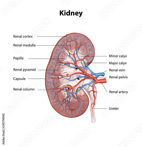
Right kidney, left kidney, fibrous capsule, renal cortex, right renal artery, right renal vein, inferior vena.
These are the functional units. Kidney function is derived from the actions of about 1.3 million nephrons per kidney; Filtered blood leaves the glomerulus via the efferent arteriole, which becomes the interlobular vein. Each medulla is composed of structures called renal pyramids. Your main goal in the kidney will be to understand how blood vessels and nephrons are organized in the kidney cortex and medulla, and how this arrangement is related to the production of urine. These are the functional units. The inner portion of each kidney contains a region called the renal medulla. This will be a smaller amount of blood, since much of the blood would leave the capillaries at the glomerulus and enter the nephron. From these arterioles branch the afferent arterioles.each afferent arteriole divides into a capillary network. Home » unlabelled » kidney blood vessels labeled / this article covers the blood supply, innervation, lymphatic drainage of the kidneys and related neurovascular supply of the kidney: Renal blood vessels labeled : This article will discuss the anatomy and major functions of the kidney. But they are depicted in most pictures and labeled as segmental veins, or not labeled.
Label the blood vessels of the kidney in figure 11.5 by filling in the blanks below the figure. Utilizing the kidney and nephron models, locate the following vessels: Labeled a in this picture The renal artery enters through the hilum, which is located where the kidney curves inward in a concave shape. Renal blood vessels labeled :
Human Kidney Cross Section Scientific Background Anatomy Urinary System With Main Parts Labeled 3d Illustration Stock Illustration Adobe Stock from as1.ftcdn.net Processed blood is removed from the kidneys and returned to circulation through blood vessels called the renal veins. Filtered blood leaves the glomerulus via the efferent arteriole, which becomes the interlobular vein. Blood supply of the kidney: The interlobar arteries which pass between the renal pyramids, arch around the base of the pyramid as the arcuate. The small artery that carries blood away from the capillaries of the glomerulus. The renal columns house blood vessels figure 24.3 internal anatomy of the kidney, including the nephron. Identify the anatomical structures of the kidney. The kidneys are important to the body's production of urine.
These give off a series of branches which enter the cortex as interlobular arterioles.
Your main goal in the kidney will be to understand how blood vessels and nephrons are organized in the kidney cortex and medulla, and how this arrangement is related to the production of urine. The renal cortex and medulla contain a complex network of blood vessels. Blood supply of the kidney: Utilizing the kidney and nephron models, locate the following vessels: This article will discuss the anatomy and major functions of the kidney. Oxygenated blood comes to the kidneys. The renal pelvis connects the kidney to the rest of the body. The dye flows through the catheter into the kidney artery. Make sure that you understand the functions of these blood vessels (use your textbook as a resource) renal arteries. They cleanse the blood of toxins and balance the constituents of the circulation to homeostatic set points through the processes of filtration, reabsorption, and secretion. This will be a smaller amount of blood, since much of the blood would leave the capillaries at the glomerulus and enter the nephron. Kidney function is derived from the actions of about 1.3 million nephrons per kidney; Processed blood is removed from the kidneys and returned to circulation through blood vessels called the renal veins.